Laser Microscope Can Diagnose Skin Cancer and Perform Surgery
The device can digitally scanning living tissue, but also treat the tissue by intensifying the laser's heat.
University of British Columbia researchers have developed a specialized microscope that has the potential ability to both diagnose diseases including skin cancer and perform incredibly precise surgery--all without cutting skin.
The researchers describe the technology in Science Advances.
"Our technology allows us to scan tissue quickly, and when we see a suspicious or abnormal cell structure, we can perform ultra-precise surgery and selectively treat the unwanted or diseased structure within the tissue--without cutting into the skin," says Yimei Huang, co-lead author of the study and a former postdoctoral fellow at the department of dermatology and skin science at UBC and BC Cancer, in a news release.
Huang co-led the study with Zhenguo Wu, a UBC PhD student.
The device is a specialized type of multiphoton excitation microscope that allows imaging of living tissue up to about one millimeter in depth using an ultrafast infrared laser beam. What sets the researchers' microscope apart from previous technology is that it's capable of not only digitally scanning living tissue, but also treating the tissue by intensifying the heat produced by the laser.
When used to treating skin diseases, the microscope allows medical professionals to pinpoint the exact location of the abnormality, diagnose it and treat it instantly. It could be used to treat any structure of the body that is reached by light and that requires extremely precise treatment, including nerves or blood vessels in the skin, eye, brain or other vital structures.
"We can alter the pathway of blood vessels without impacting any of the surrounding vessels or tissues," says study co-author Harvey Lui, professor at the department of dermatology and skin science at UBC and the Vancouver Coastal Health Research Institute, and a dermatologist at BC Cancer. "For diagnosing and scanning diseases like skin cancer, this could be revolutionary."
The researchers wanted to make multiphoton microscope technology more versatile while also increasing its precision.
"We wanted to be able to identify what was happening under the skin from many different angles and to have the capability of imaging different body sites," adds senior author Haishan Zeng, professor of dermatology, pathology and physics at UBC and distinguished scientist with BC Cancer.
"Once we achieved that, we wondered whether we could transform this diagnostic device into a treatment device by simply turning up the power of the laser."
The results were incredibly exciting.
"We are not only the first to achieve fast video-rate imaging that enables clinical applications, but also the first to develop this technology for therapeutic uses," says Zeng.
The researchers have partnered with several UBC departments, including mechanical engineering, electrical engineering and ophthalmology, to develop different versions of the technology. Exploration includes research into the development of a miniature version that could be used to perform microscopic examinations and treatment during endoscopy--a non-surgical procedure used to examine a person's digestive tract using an endoscope, a flexible tube with a light and camera attached to it.
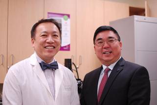
IMAGE: FROM LEFT TO RIGHT: CO-AUTHOR HARVEY LUI AND SENIOR AUTHOR HAISHAN ZENG
CREDIT: UBC
Facebook Comments