
Cherry Angiomas Linked to NF1-Related Vascular Changes: Study
Key Takeaways
- Cherry angiomas were more common among individuals with neurofibromatosis type 1 (NF1) than controls (48% vs 18%), according to new research.
- Somatic NF1 loss-of-function second-hit variants were identified in 67% of NF1-associated cherry angiomas in the study.
- Molecular analyses localized NF1 second hits predominantly to endothelial cells and telocytes and showed increased MAPK pathway signaling in these vascular cell populations.
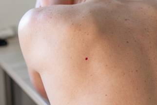
Cherry angiomas may be a previously underrecognized vascular manifestation of neurofibromatosis type 1 (NF1), according to findings from a prospective comparative study in JAMA Dermatology.
Investigators evaluated 259 participants (102 individuals with confirmed NF1 and 157 controls without NF1) from 2020 to 2021 in a single French national referral center for neurofibromatoses at Henri-Mondor University Hospital in Créteil, France. Study participants were aged 15 years or older.
Cherry Angiomas Were More Common and Occurred Earlier in NF1
Cherry angiomas were identified in 48% of participants with NF1 compared with 18% of controls, corresponding to an odds ratio of 4.26 (95% CI, 2.44 to 7.56). The lesions also occurred at younger ages among individuals with NF1. The association persisted after adjustment for age and sex and in propensity score–matched analyses. Somatic NF1 loss-of-function second-hit variants were detected in 26 of 39 NF1-associated cherry angiomas (67%) but in none of the control lesions.
Cell-specific sequencing localized NF1 second hits primarily to endothelial cells and telocytes, with higher variant allele frequencies in endothelial cells. Immunofluorescence demonstrated increased phospho–extracellular signal-regulated kinase signaling in these vascular cell populations, providing evidence of rat sarcoma–mitogen-activated protein kinase pathway activation.
“The results of this cross-sectional study suggest that CAs represent a frequent and previously unrecognized vascular manifestation of NF1,” the authors wrote in the study. “These findings potentially expand the spectrum of NF1-associated neoplasms and establish CAs as a model for NF1-related vasculopathy.”
Source
Fertitta L, et al. JAMA Dermatology. 2026. Doi:10.1001/jamadermatol.2026.2933