
Evidence, Techniques, and a Standardized 5-Fluorouracil Protocol for Intralesional Therapies Treating Keratinocyte Cancers
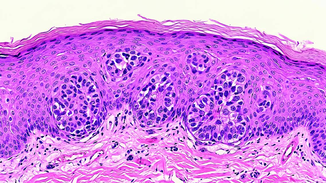
The standard treatment for squamous cell carcinoma (SCC), keratoacanthoma (KA), and basal cell carcinoma (BCC) is surgical. With the increasing incidence of keratinocyte cancer (KC), intralesional therapies present an effective alternative therapeutic modality.¹ Despite decades of use and reported efficacy, intralesional (IL) therapies remain underutilized in routine clinical practice, in part due to variability in clinician familiarity, limited high-level evidence, and the absence of standardized treatment protocols. Intralesional therapy has been used to treat keratinocyte tumors (including KA) for decades, with agents such as 5-fluorouracil (5-FU), methotrexate (MTX), interferons, and bleomycin (Table 1).2,3 This treatment approach is of particular interest for patients who are not surgical candidates, are interested in non-surgical treatments, have a high tumor burden, have tumors in challenging locations, or face financial limitations.4 The use of IL therapies has also been reported to be beneficial in cases of post-procedural tumor recurrence (particularly for KA).2,5 Some challenges to the use of IL therapies include off-label use of agents, the retrospective nature of many studies, local and systemic risks of therapy, and significant variability in dosing, administration techniques, and treatment intervals across studies, which has limited broader adoption and standardization of care.6-8

INTRALESIONAL FLUOROURACIL
5-FU is an antimetabolite agent that has previously been used as a systemic therapy in the treatment of cutaneous and internal malignancies.² This agent has been reported to be an effective treatment for KA, SCC, and BCC,2,4-6,9,10 with particular efficacy observed in rapidly proliferating lesions such as KA, consistent with its preferential activity in tissues with high mitotic rates.² Parker and Hanke reported the use of intralesional 5-FU (IL5-FU) in the treatment of five large (>20 mm) KA on the face, of which four (80%) were cleared without recurrence at follow-up (18–45 months).² All participants underwent laboratory evaluation, including blood counts and chemistry panels, prior to injections, and none demonstrated treatment-related changes. Parker and Hanke theorized that IL5-FU may be less effective in KA that has undergone an intrinsic halt in rapid cellular proliferation.² n
Que, Compton, and Schmults reported the use of IL5-FU as an effective treatment for eruptive KA in a cohort of 30 participants, including immunosuppressed patients; there were no lesion recurrences after treatment and a reported 90% decrease in the mean number of surgical excisions required on the IL5-FU–treated extremity.5 Hamad et al reported successful clearance of 89% of 315 keratinocyte tumors in 61 patients treated with debulking followed by IL5-FU, including SCC in situ, invasive SCC, SCC (KA-type), and KA. Factors associated with treatment failure included immunosuppressed status, head and neck location, and large tumor size (>19 mm).⁴ Some studies have also reported clearance of BCC using IL5-FU therapy, IL5-FU/epinephrine gel, and sustained-release IL5-FU/epinephrine implants.4,6,9,10 Additional studies have reported cure rates for KC ranging from 95% to 100%.7,8,11 While these outcomes are promising, the majority of available data are derived from retrospective case series and small cohort studies, which may limit generalizability.
Reported side effects of IL5-FU include injection-site reactions such as dyspigmentation, erosions and/or ulceration, temporary alopecia, and minimal to no systemic effects (eg, mild nausea—with one case requiring antiemetics—subjective fatigue, and headache).2,4,5,9,10 Most reports describe the use of lidocaine with epinephrine for local anesthesia prior to IL5-FU administration due to associated pain, with the added potential benefit of increased efficacy from epinephrine-induced vasoconstriction.
Compared with other intralesional agents discussed below, 5-FU represents an accessible, cost-effective option, with an average cost ranging from $12 to $26 for a 50 mL vial, and relatively fewer systemic side effects.6-8 However, a key limitation of IL5-FU is the significant variability in administration techniques and dosing regimens across studies. Commonly reported regimens include a 50-mg/mL concentration of 5-FU, injection volumes ranging from 0.1–3 mL per treatment, treatment intervals of every 2–4 weeks, and the need for multiple treatment sessions.2,4-8,10 This heterogeneity has contributed to inconsistent adoption in clinical practice and highlights the need for more standardized approaches. To address this limitation, we include in this report a standardized IL5-FU protocol utilized at our institution (Table 2).

INTRALESIONAL METHOTREXATE
Methotrexate (MTX) is an antifolate agent that has been reported for use in treating BCC, KA, and SCC.7,8,11 Similar to other intralesional antimetabolites, its efficacy is thought to be related to inhibition of DNA synthesis in rapidly proliferating tumor cells. Reported cure rates for keratinocyte tumors range from 83% to 100%,7,8,11,12 although the available evidence is primarily limited to small retrospective series and cohort studies.
Clinical reports support its use across a range of keratinocyte tumors. Intralesional MTX (ILMTX) dosing typically ranges from 0.2–2 mL of 12.5- or 25-mg/mL concentrations,7,8,11-13 with injections administered at approximately 2-week intervals for an average of one to two treatment sessions. Reported advantages compared with IL5-FU include the potential to avoid local anesthesia and, in some cases, a reduced number of treatment visits.11 In addition, a combination approach using tumor debulking followed by ILMTX has demonstrated high efficacy, with reported clearance rates of up to 95%.13
Reported adverse effects are generally localized and include injection-site reactions. Systemic toxicity is uncommon but has been described, particularly in patients with renal impairment, and may include hematologic abnormalities (ranging from anemia to pancytopenia), mucositis, epistaxis, malaise, fatigue, and hepatotoxicity.8,11-15
Compared with other intralesional agents, ILMTX is a relatively accessible and low-cost option, with an average reported cost ranging from $21.39–$25 for 250 mg in 10 mL.7,8
A limitation of ILMTX, similar to other intralesional therapies, is variability in dosing protocols and patient selection across studies, which has contributed to heterogeneity in reported outcomes and limits direct comparison with surgical standards of care.
INTRALESIONAL BLEOMYCIN
Bleomycin is an antineoplastic agent that exerts cytotoxic effects through DNA strand breaks during the cell cycle and has been used intralesionally for the treatment of BCC, KA, and SCC in situ.8,11,16 However, compared with other intralesional therapies, its use in keratinocyte carcinomas is less common, and the strongest evidence base for IL bleomycin remains in the treatment of viral warts rather than skin cancer.
When used for keratinocyte tumors, reported clearance rates range from 98% to 100%,7,8,11,17,18 although these outcomes are largely derived from small case series and isolated reports, limiting generalizability. IL administration is typically performed using doses ranging from 0.2–1.5 IU, adjusted based on tumor size and treatment frequency.7,8,11,17,18
To enhance tissue penetration and therapeutic effect, some reports describe the use of electroporation-assisted delivery, which transiently increases cell membrane permeability to improve drug uptake.¹7 In addition, a single case report describes the use of a chalazion clamp to localize therapy for auricular BCC, highlighting procedural adaptations for difficult anatomical sites.18
Reported adverse effects include localized injection-site reactions, infection, and muscle contractions, particularly in the setting of electroporation-assisted delivery. Compared with other intralesional agents, bleomycin is associated with a wider cost range, with reported average costs of $26.14 to $350 per 15 mg via.3,7,8,11,19,20
A key limitation of IL bleomycin is the relative paucity of high-quality data in keratinocyte carcinomas and its more established role in the treatment of warts, which has contributed to its more limited adoption in cutaneous oncology practice.
INTRALESIONAL INTERFERON
Interferons (IFN) are cytokines with antiproliferative, immunomodulatory, and antiviral properties that have been used as intralesional treatments for non-melanoma skin cancers (NMSC). Reported formulations include IFN alpha-2a, IFN alpha-2b, IFN beta, and IFN gamma.3,7,8,11,19,20 Intralesional IFNs have been described as treatment options for keratoacanthoma (KA), squamous cell carcinoma (SCC), and basal cell carcinoma (BCC), although most available data support the use of IFN alpha, particularly for BCC and KA.3,7,8,11,19,20
Reported efficacy is highly variable and appears to depend on interferon subtype. IFN alpha-2a and alpha-2b demonstrate the widest range of reported response rates (0%–100%), IFN beta ranges from approximately 50%–67%, and IFN gamma from 0%–50%.3,7,8,11,19,20 This variability reflects the heterogeneity of available studies, which are largely limited to small case series and early clinical reports.
Treatment regimens have varied considerably, with reported dosing ranging from 0.2–30 million IU administered three times weekly for approximately 3 weeks, though a commonly used dose is 1.5 million IU.3,7,8,11,19,20
Reported adverse effects are systemic and dose-limiting, and include injection-site reactions, flu-like symptoms (fever, chills, myalgias, arthralgias), headache, gastrointestinal symptoms, back pain, rigors, depressive symptoms, neutropenia, and occasional alterations in liver enzymes and blood counts.19,20
Compared with other intralesional agents, IFNs are associated with substantially higher cost, with reported average treatment costs ranging from $166.23 to $3,287,13 depending on interferon subtype and dosing regimen.7,8
A major limitation of intralesional IFN therapy is the marked variability in reported efficacy, high cost, and systemic side effect profile, which together have limited its widespread adoption in current dermatologic practice.
OTHER INTRALESIONAL THERAPIES ON THE HORIZON
Talimogene laherparepvec (T-VEC) is an oncolytic virus therapy derived from a modified herpes simplex virus type 1 (HSV-1) that is FDA approved for the treatment of melanoma.21,22 It has also been investigated for the treatment of non-melanoma skin cancers, with reported clearance rates of approximately 90% in low-risk cutaneous squamous cell carcinoma (SCC)²¹. The reported dosing protocol includes an initial dose of 1010 PFU/mL followed by 10⁸ PFU/mL for up to 4 intralesional injections administered every 2 weeks.²¹ Adverse effects are generally mild to moderate and include flu-like symptoms, headache, and fatigue, typically resolving within 1–2 days.²¹
Cemiplimab is a programmed death-1 (PD-1) inhibitor that confers antitumor effects through immune checkpoint blockade. It is currently FDA approved for the treatment of locally advanced or metastatic cutaneous SCC and locally advanced or metastatic BCC. Intralesional administration is currently being investigated in early-stage SCC through ongoing clinical trials (NCT06585410), reflecting growing interest in localized immune checkpoint therapy.
Although both T-VEC and intralesional immunotherapy with agents such as cemiplimab represent promising advances in the field, their clinical utility is currently limited by cost, which is substantially higher than traditional intralesional agents reviewed in this manuscript, as well as by limited data in early-stage keratinocyte carcinomas.
CONCLUSION
Keratinocyte cancers (KC) are most effectively treated with surgical intervention; however, intralesional therapies represent a valuable and increasingly supported alternative for appropriately selected patients, particularly those who are not surgical candidates or who prefer nonsurgical management. Across available studies, intralesional therapies demonstrate generally high efficacy, with improved outcomes observed for tumors located on the trunk and extremities compared with those on the head and neck.
Selection of an intralesional agent should be individualized, incorporating tumor characteristics (including size, location, and recurrence risk), patient-specific factors (such as comorbidities and immune status), potential adverse effects, treatment burden, and overall cost. Traditional agents such as 5-FU and MTX remain cost-effective and accessible options with favorable safety profiles, while emerging therapies, including oncolytic viral and immune-based approaches, demonstrate promising efficacy but are currently limited by cost, availability, and lack of extensive real-world data in early-stage disease.
Overall, intralesional therapies expand the therapeutic armamentarium for KC and provide a practical, tissue-sparing alternative in carefully selected clinical scenarios. As evidence continues to evolve, future prospective studies and efforts toward standardization of dosing and administration will be essential to better define their role within contemporary skin cancer management.
Disclosures: The authors report no relevant disclosures.
1. Zhou L, Zhong Y, Han L, Xie Y, Wan M. Global, regional, and national trends in the burden of melanoma and non-melanoma skin cancer: insights from the global burden of disease study 1990-2021. Sci Rep. 2025;15(1):5996. Published 2025 Feb 18. doi:10.1038/s41598-025-90485-3
2. Parker CM, Hanke CW. Large keratoacanthomas in difficult locations treated with intralesional 5-fluorouracil. J Am Acad Dermatol. 1986;14(5 Pt 1):770-777. doi:10.1016/s0190-9622(86)70092-3
3. Grob JJ, Suzini F, Richard MA, et al. Large keratoacanthomas treated with intralesional interferon alfa-2a. J Am Acad Dermatol. 1993;29(2 Pt 1):237-241. doi:10.1016/0190-9622(93)70174-r
4. Hamad J, Jolly PS. Debulking followed by intralesional 5-fluorouracil for the treatment of cutaneous squamous cell carcinoma and keratoacanthoma: A retrospective analysis. Dermatol Ther. 2021;34(6):e15139. doi:10.1111/dth.15139
5. Que SKT, Compton LA, Schmults CD. Eruptive squamous atypia (also known as eruptive keratoacanthoma): Definition of the disease entity and successful management via intralesional 5-fluorouracil. J Am Acad Dermatol. 2019;81(1):111-122. doi:10.1016/j.jaad.2018.10.014
6. Metterle L, Nelson C, Patel N. Intralesional 5-fluorouracil (FU) as a treatment for nonmelanoma skin cancer (NMSC): A review. J Am Acad Dermatol. 2016;74(3):552-557. doi:10.1016/j.jaad.2015.09.040
7. Chitwood K, Etzkorn J, Cohen G. Topical and intralesional treatment of nonmelanoma skin cancer: efficacy and cost comparisons. Dermatol Surg. 2013;39(9):1306-1316. doi:10.1111/dsu.12300
8. Kirby JS, Miller CJ. Intralesional chemotherapy for nonmelanoma skin cancer: a practical review. J Am Acad Dermatol. 2010;63(4):689-702. doi:10.1016/j.jaad.2009.09.048
9. Miller BH, Shavin JS, Cognetta A, et al. Nonsurgical treatment of basal cell carcinomas with intralesional 5-fluorouracil/epinephrine injectable gel. J Am Acad Dermatol. 1997;36(1):72-77. doi:10.1016/s0190-9622(97)70328-1
10. Orenberg EK, Miller BH, Greenway HT, et al. The effect of intralesional 5-fluorouracil therapeutic implant (MPI 5003) for treatment of basal cell carcinoma. J Am Acad Dermatol. 1992;27(5 Pt 1):723-728. doi:10.1016/0190-9622(92)70245-b
11. Good LM, Miller MD, High WA. Intralesional agents in the management of cutaneous malignancy: a review. J Am Acad Dermatol. 2011;64(2):413-422. doi:10.1016/j.jaad.2009.12.013
12. Moss M, Weber E, Hoverson K, Montemarano AD. Management of Keratoacanthoma: 157 Tumors Treated With Surgery or Intralesional Methotrexate. Dermatol Surg. 2019;45(7):877-883. doi:10.1097/DSS.0000000000001739
13. Chandawarkar SK, Surapaneni N, Oliver C, et al. Retrospective cohort of 168 keratinocytic tumors treated with combination therapy of surgical debulking followed by intralesional methotrexate. J Am Acad Dermatol. Published online February 25, 2026. doi:10.1016/j.jaad.2026.02.081
14. Lamberg O, Powers JG, Negbenebor NA. A case of systemic toxicity after intralesional methotrexate for non-melanoma skin cancer in a patient with chronic kidney disease. JAAD Case Rep. 2026;70:4-6. Published 2026 Feb 2. doi:10.1016/j.jdcr.2026.01.039
15. Flynn KN, Johnson MS, Brink WC, Smith DL. Pancytopenia, mucositis, and hepatotoxicity after intralesional methotrexate injection in a patient treated with peritoneal dialysis. Am J Health Syst Pharm. 2012;69(7):578-582. doi:10.2146/ajhp110252
16. Bik L, Sangers T, Greveling K, Prens E, Haedersdal M, van Doorn M. Efficacy and tolerability of intralesional bleomycin in dermatology: A systematic review. J Am Acad Dermatol. 2020;83(3):888-903. doi:10.1016/j.jaad.2020.02.018
17. Glass LF, Jaroszeski M, Gilbert R, Reintgen DS, Heller R. Intralesional bleomycin-mediated electrochemotherapy in 20 patients with basal cell carcinoma. J Am Acad Dermatol. 1997;37(4):596-599. doi:10.1016/s0190-9622(97)70178-6
18. Eikenberg JD, Grider DJ, Kolodney MS. Use of clamping to enhance intralesional bleomycin therapy for nodular basal cell carcinoma. JAAD Case Rep. 2014;1(1):7-8. Published 2014 Sep 19. doi:10.1016/j.jdcr.2014.08.001
19. Oh CK, Son HS, Lee JB, Jang HS, Kwon KS. Intralesional interferon alfa-2b treatment of keratoacanthomas. J Am Acad Dermatol. 2004;51(5 Suppl):S177-S180. doi:10.1016/j.jaad.2004.05.009
20. Edwards L, Whiting D, Rogers D, Luck K, Smiles KA. The effect of intralesional interferon gamma on basal cell carcinomas. J Am Acad Dermatol. 1990;22(3):496-500. doi:10.1016/0190-9622(90)70070-x
21. Stratton DB, Adams AC, Sundararajan S, Cui H, Roe DJ, Curiel-Lewandrowski C. A single-arm phase 2 study of talimogene laherparepvec in patients with lower-risk invasive cutaneous squamous cell cancer. J Am Acad Dermatol. 2026;94(3):859-866. doi:10.1016/j.jaad.2025.11.026
22. Stull CM, Clark D, Parker T, Idriss MH, Patel VA, Migden MR. Current and emerging intralesional immunotherapies in cutaneous oncology. J Am Acad Dermatol. 2024;91(5):910-921. doi:10.1016/j.jaad.2024.05.095
Puneet Singh Jolly, MD
- Professor of Dermatology
- University of North Carolina, Department of Dermatology
Chapel Hill, N.C.
Maria D. Melendez-Gonzalez, MD
- Assistant Professor of Dermatology
- University of North Carolina, Department of Dermatology
Chapel Hill, N.C.
Ready to Claim Your Credits?
You have attempts to pass this post-test. Take your time and review carefully before submitting.
Good luck!
Recommended
 Skin Cancer & Photoprotection
Mohs Micrographic Surgery Access and Distribution Lacking in Louisiana
Randy Head II, BS; Amanda Porter, MDRandy Head II, BS; Amanda Porter, MD- Skin Cancer & Photoprotection
Advanced Management Solutions for High-Grade Cutaneous Toxicities to Cancer Therapy
Preetha Muralidharan, MBBS, MPH; Rebeca Martinez, MDPreetha Muralidharan, MBBS, MPH; Rebeca Martinez, MD - Skin Cancer & Photoprotection
Tips for Management of Cutaneous Squamous Cell Carcinoma
Amanda Stines, CCMA; Vishal A. Patel, MD, FAADAmanda Stines, CCMA; Vishal A. Patel, MD, FAAD