
Severe Widespread Eczema Mimicking Mycosis Fungoides
Mycosis fungoides, the most common subtype of cutaneous T-cell lymphoma, is well recognized for its ability to mimic a wide array of benign inflammatory dermatoses.
KEY TAKEAWAYS
- Severe adult atopic dermatitis (AD) can closely mimic mycosis fungoides, especially when presenting with widespread, indurated, violaceous, or lichenified plaques and atypical distribution.
- Clinicopathologic correlation is critical, as substantial clinical and histopathologic overlap exists between the two conditions.
- Histopathology, immunohistochemistry, and T-cell receptor gene rearrangement studies can help clarify the diagnosis, and ancillary testing should be interpreted alongside clinical findings and longitudinal assessment.
- Psychological stress may substantially exacerbate AD and alter its clinical presentation, reminding dermatologists to consider psychosocial and environmental factors when evaluating severe or atypical eczematous eruptions.
Atopic dermatitis (AD) is a chronic inflammatory dermatosis characterized by pruritus, erythema, epidermal barrier dysfunction, and immune dysregulation. Although traditionally considered a disease of childhood, AD frequently persists into adulthood or presents de novo later in life, often with atypical morphology and distribution.1 Adult disease may manifest with lichenified plaques, erythroderma, or widespread indurated lesions that challenge diagnostic certainty and broaden the differential diagnosis to include inflammatory, infectious, and neoplastic processes.2
Mycosis fungoides, the most common subtype of cutaneous T-cell lymphoma, is well recognized for its ability to mimic a wide array of benign inflammatory dermatoses. Early-stage disease often presents with nonspecific patches or plaques accompanied by pruritus and subtle scale, which can lead to delays in diagnosis. Multiple reviews have described mycosis fungoides as a clinical mimic of other conditions, with AD among the most frequently noted conditions both clinically and histopathologically.3-5 This overlap can result in significant diagnostic uncertainty, particularly in young adults without classic risk factors for lymphoma.
Psychological stress is an established exacerbating factor in inflammatory skin disease. Neuroimmune interactions involving the hypothalamic-pituitary-adrenal axis and sympathetic nervous system contribute to altered cortisol responses, cytokine release, and impairment of epidermal barrier function.6 Such stress can intensify or modify the clinical expression of AD. These stress-related effects may further obscure the distinction between inflammatory dermatoses and early cutaneous lymphoma.
We present a case of severe, widespread AD in a young patient whose clinical presentation closely mimicked mycosis fungoides, which prompted extensive diagnostic evaluation. This case underscores the importance of clinicopathologic correlation and longitudinal assessment when evaluating atypical or severe eczematous eruptions.
CASE PRESENTATION
A 24-year-old man presented with a rapidly progressive, intensely pruritic eruption involving the trunk and proximal extremities. The eruption developed over several weeks during a period of marked psychological stress related to preparation for professional licensing examinations. The patient reported severe anxiety, insomnia, and prolonged outdoor activity with extensive sun exposure. He denied fever, night sweats, unintended weight loss, or systemic symptoms. The patient’s medical history was notable for alopecia areata diagnosed in childhood. The family history was significant for autoimmune disease in first-degree relatives. He reported a history of childhood eczema that had been quiescent for several years before the current flare.
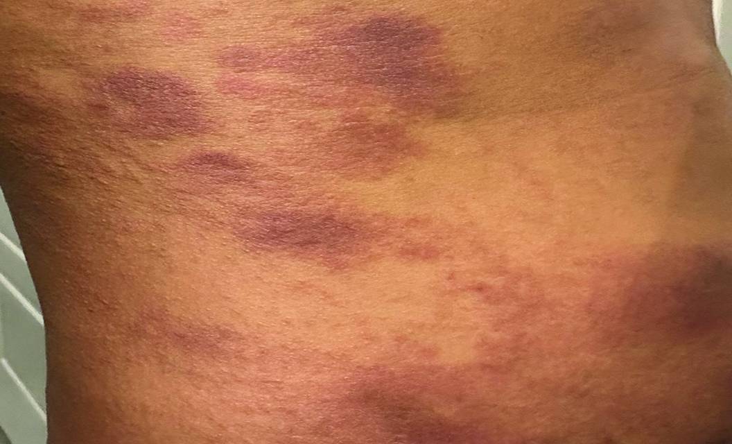
Cutaneous examination revealed widespread erythematous to violaceous plaques with irregular borders and variable induration. Lesions demonstrated areas of lichenification and fine scale, with heterogeneous morphology and poor demarcation, involving the posterior trunk (Figure 1), lateral abdomen (Figure 2), and anterior thigh (Figure 3). The distribution and plaque quality raised concern for a lymphoproliferative disorder.

Figure 1.

Figure 2.

Figure 3.
Given the chronicity, morphology, and distribution of lesions, mycosis fungoides was strongly considered. Multiple punch biopsies were obtained from representative sites on the trunk and thigh. Histopathologic examination demonstrated spongiotic dermatitis with acanthosis, parakeratosis, and a superficial perivascular lymphocytic infiltrate. No significant epidermotropism, atypical lymphocytes, or Pautrier microabscesses were identified. Immunohistochemical staining revealed preserved pan T-cell markers, and molecular analysis did not demonstrate a clonal T-cell receptor gene rearrangement. These findings supported a diagnosis of severe eczematous dermatitis rather than cutaneous T-cell lymphoma.
The patient was treated with intralesional triamcinolone to the most indurated plaques, a short tapering course of oral prednisone, and high-potency topical corticosteroids, along with liberal use of emollients. Stress reduction strategies were discussed, including sleep optimization and modification of daily routines. At the 3-week follow-up, the patient demonstrated marked clinical improvement, with a substantial reduction in erythema, plaque thickness, and body surface area involvement.
DISCUSSION
This case illustrates the substantial clinical and histopathologic overlap between severe AD and early mycosis fungoides. AD is increasingly recognized as a systemic inflammatory disorder involving Th2-skewed immune responses, elevated cytokine production, and epidermal barrier dysfunction. In adults, disease expression is often atypical, with lichenified or indurated plaques that may resemble inflammatory or neoplastic dermatoses rather than classic flexural eczema.2
Mycosis fungoides exhibits marked heterogeneity in its early stages, frequently lacking pathognomonic features. Patches and plaques may be poorly demarcated, pruritic, and scaly, closely resembling chronic dermatitis. Diagnostic ambiguity is compounded by the fact that inflammatory dermatoses may demonstrate reactive lymphoid infiltrates that mimic lymphoma.
Histopathologic overlap remains a major challenge. Spongiosis, acanthosis, and superficial perivascular lymphocytic infiltrates may be present in both entities. Whereas epidermotropism of atypical lymphocytes, cytologic atypia, and loss of pan T-cell markers favor mycosis fungoides, these features may be subtle or absent in early disease. Ancillary studies, including immunohistochemistry and T-cell receptor gene rearrangement analysis, improve diagnostic accuracy but are not definitive in isolation.7 Likewise, biomarker studies do not provide a reliable, noninvasive method to distinguish mycosis fungoides from AD, as cytokine profiles often demonstrate substantial overlap.8 Recently, dermoscopy has emerged as a useful adjunctive tool in distinguishing early mycosis fungoides from inflammatory dermatoses. Although considerable overlap persists, differences in vascular morphology and scaling patterns among mycosis fungoides, AD, and psoriasis have been identified.9
Psychological stress likely played a central role in exacerbating this patient’s disease presentation. The patient’s recent pressure and lifestyle associated with licensing examination preparation could have intensified his clinical manifestation. Furthermore, his autoimmune history of alopecia areata supports an underlying predisposition to such an eruption.
CONCLUSION
Severe AD in adults can present with atypical morphology that closely resembles cutaneous T-cell lymphoma, posing a significant diagnostic challenge. In this case, the combination of indurated, violaceous plaques and widespread distribution warranted thorough evaluation, including histopathology, immunohistochemistry, and molecular studies, to exclude mycosis fungoides. The role of psychological stress as an exacerbating factor further underscores the need to consider clinical context when interpreting ambiguous presentations. This case highlights the importance of a broad differential diagnosis and careful clinical approach to differentiate mycosis fungoides from common inflammatory dermatoses, such as eczema. Clinicopathologic correlation, ancillary testing, and longitudinal follow-up remain essential in ensuring diagnostic accuracy and guiding appropriate management.
Ready to Claim Your Credits?
You have attempts to pass this post-test. Take your time and review carefully before submitting.
Good luck!
Recommended
 Atopic Dermatitis
Nonsteroidal Topical Therapies for Atopic Dermatitis from Infants to Adults
Alice Tang, BA; Benjamin Ungar, MDAlice Tang, BA; Benjamin Ungar, MD- Atopic Dermatitis
The Importance of Patient-Reported Outcomes in Atopic Dermatitis
Aaron Cheng, BA, BS; David CotterAaron Cheng, BA, BS; David Cotter