PracticalDermatology

Conjunctival Squamous Cell Carcinoma: A Video-Illustration of a Surgical Approach
Squamous cell carcinoma (SCC) of the skin is an invasive carcinoma of the epidermis. This malignancy arises most often in areas with chronic sun exposure, such as the forehead, ears, and hands. Clinically, cutaneous SCC typically consists of an erythematous skin ulcer surrounded by a raised, indurated border. On histopathologic examination, the tumor is characterized by the irregular proliferation of atypical squamous cells downward through the basement membrane into the dermis. The pathognomonic morphology of squamous cell carcinoma is cellular atypia extending through the full thickness of the epidermis.1 Cellular atypia specifically describes the variation of the size and shape of cells and nuclei, the absence of intercellular bridges, and the presence of atypical mitotic figures.1
Squamous cell carcinoma can also arise within mucocutaneous surfaces, including the conjunctiva.2,3 The conjunctiva is a unique mucous membrane that covers the anterior surface of the eye and is partially exposed to sunlight.3 In the conjunctiva, particularly in the sun-exposed areas nasally and temporally, SCC can develop and appears as a gelatinous, vascular, or leukoplakic mass typically located at the corneoscleral limbus. Histopathologic examination can reveal a localized lesion that remains confined to the surface epithelium, known as conjunctival intraepithelial neoplasia or dysplasia (CIN), or invasive SCC with malignant cells that grow in sheets and cords through the basement membrane into the deeper stroma. While CIN has no metastatic potential, SCC of the conjunctiva can enter the lymphatics and metastasize to local lymph nodes.3
Several studies over the past decades have attempted to quantify metastatic rates of cutaneous squamous cell carcinoma, arriving at rates of approximately 1.3 percent to 3.7 percent.2,4-6 Larger tumor dimension and poorly differentiated tumors are independent predictors for nodal metastasis and disease-specific death.5
Metastasis of conjunctival SCC is uncommon, though immunosuppressed patients are at greater risk for metastatic disease. Management of conjunctival SCC depends on the location and extent of the lesion.3 Herein, we describe a patient with conjunctival SCC and video-illustrate the surgical approach. The video can be viewed online at PracDerm.com/ConjunctivalSCC.
Case Report
A 68-year-old white male with a history of basal cell carcinoma of the right lower eyelid, surgically excised 10 years prior to presentation, was referred to the Ocular Oncology Service at Wills Eye Hospital, Philadelphia for an asymptomatic conjunctival lesion on the right eye.
On our examination, best-corrected visual acuity was 20/30 in the right eye and 20/20 in the left eye. The left eye was unremarkable. Evaluation of the right eye revealed a gelatinous, vascular conjunctival mass temporally at the limbus, measuring 13mm in diameter and 3mm in thickness. The lesion demonstrated several feeder blood vessels from the fornix and with classic “hairpin loops.” There was no intraocular invasion, and the tarsal and forniceal conjunctiva were clean. These features were characteristic of conjunctival SCC.
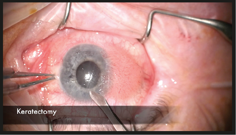
The patient was treated with “no-touch” surgical excision beginning with absolute alcohol applied to the localized corneal surface for epithelial keratectomy, cauterization of feeder vessels, and dissection of the tumor off the globe with 2-3mm margins, and placement of tumor in fixative for histopathology. Using clean instruments, conjunctival “double freeze-thaw” cryotherapy and reconstruction with conjunctivoplasty and double layer closure was performed. Tissue glue and corticosteroid/antibiotic ointment was applied following closure. Histopathology of the specimen demonstrated an abrupt transition from unremarkable conjunctival epithelium into a papillomatous configuration with acanthosis, parakeratosis, and dysplastic epithelium, consistent with conjunctival SCC. Koilocytosis was also noted, suggesting a human papillomavirus cytopathic effect. The patient healed well by three weeks (Video at 2:58) without recurrence at one-year follow-up.
WATCH NOW

“No-touch” surgical excision and “double freeze-thaw” cryotherapy techniques.
Guide: 00:11: Keratectomy; 00:18: Hemostasis with bipolar cautery; 00:23: Careful resection with minimal manipulation (“no-touch technique”); 00:49: Careful dissection of tumor off sclera; 01:02: Dissection of tumor off limbus; 01:30: Tumor placed on cardboard and floated in fixative for pathology; 01:32: Hemostasis with bipolar cautery; 01:41: Clean-up of the scleral surface with a 57 scleral blade; 01:47: Lamellar scleral dissection for clean margin at the base; 01:59: Double freeze-thaw cryotherapy to ensure all conjunctival margins clean; 02:12: Ocular surface rewetting; 02:15: Conjunctivoplasty with 7.0 Vicryl suture; 02:30: Ocular surface rewetting; 02:33: Conjunctivoplasty with 7.0 Vicryl suture; 02:46: Limbal relaxation; 02:49: Application of tisseal glue; 02:54: Preoperative slit lamp photo ; 02:57: Post-operative slit lamp photo
Visit: PracDerm.com/ConjunctivalSCC
Discussion
Conjunctival SCC is one of the more common conjunctival malignancies, typically found in the older, white male population, particularly those with extensive sun exposure.7,8 Ultraviolet B light is thought to cause DNA damage of the conjunctival epithelial cells, mainly those at the sun-exposed nasal and temporal limbus, that eventually leads to mutations predisposing to development of malignant cells.7 Other known risk factors for conjunctival SCC include human papillomavirus (HPV) 16/18 infection, cigarette smoking, and ocular surface injury.7 Shields, et al. provided a comprehensive evaluation of 5,002 conjunctival tumors and found that SCC represented nine percent of the entire group.8 In that series, conjunctival SCC appeared at mean age of 67 years and with a predominance for Caucasian race (87 percent) and male sex (73 percent).8
Management of conjunctival SCC involves both surgical and nonsurgical methods. Regarding surgical management, the “no-touch” method is preferred, in which the tumor is never directly manipulated during surgery and handling of the mass is limited to only the normal conjunctiva bordering the mass. This can prevent or minimize tumor seeding into adjacent structures. Following complete tumor removal, clean (uncontaminated) instruments are used to apply cryotherapy using the “double freeze-thaw” technique, followed by the closure of Tenon’s fascia (for wound strength) and then conjunctiva (for wound cosmesis).9
Nonsurgical methods for control of conjunctival squamous neoplasia include topical chemotherapy (mitomycin C, 0.01%-0.04%/1cc or 5-fluorouracil, 0.5%-1.0%/cc), topical interferon alpha 2-b (1 million international units (IU)/cc), or injection of interferon alpha-2b (10 million IU/cc).10 Mitomycin C and 5-fluorouracil are typically used for a one-week period and can be toxic to the corneal epithelium, leading to symptomatic epitheliopathy, limbal stem cell loss, and potential corneal scarring with vision loss.11,12 Interferon is a well-tolerated topical agent for conjunctival SCC with minimal side effect profile. Interferon can be used as immunotherapy to eradicate a tumor, as immunoreduction of “giant” squamous neoplasia so that they become surgically resectable, or as immunoprevention in patients with conditions that predispose to recurrence, such as HIV infection.13 This medication works more slowly, requiring three to six months of therapy.
In summary, we describe and video-illustrate the surgical management of conjunctival SCC. Similar to its cutaneous counterpart, this lesion occurs most often in elderly Caucasian males, particularly those with extensive sun exposure.
Support provided in part by the Eye Tumor Research Foundation, Philadelphia, PA (CLS). The funders had no role in the design and conduct of the study, in the collection, analysis and interpretation of the data, and in the preparation, review or approval of the manuscript. Carol L. Shields, M.D. has had full access to all the data in the study and takes responsibility for the integrity of the data and the accuracy of the data analysis. No conflicting relationship exists for any author.
1. Smoller BR. Squamous cell carcinoma: from precursor lesions to high-risk variants. Modern Pathology. 2006;19:S88-92.
2. Weinberg AS, Ogle CA, Shim EK. Metastatic cutaneous squamous cell carcinoma: an update. Dermatol Surg 2007;33:885-99.
3. Shields CL, Shields JA. Tumors of the conjunctiva and cornea. Indian J Ophthalmol 2019;67:1930-48.
4. Khan K, Mykula R, Kerstein R, et al. A 5-year follow-up study of 633 cutaneous SCC excisions: rates of local recurrence and lymph node metastasis. Journal of Plastic, Reconstructive and Aesthetic Surgery 2018;71:1153-8
5. Schmults CD, Karia PS, Carter JB, Han J, Qureshi AA. Factors predictive of recurrence and death from cutaneous squamous cell carcinoma: a 10-year single-institution cohort study. JAMA Dermatol 2013;149(5):541-7.
6. Brantsch KD, Meisner C, Schonfisch B, et al. Analysis of risk factors determining prognosis of cutaneous squamous-cell carcinoma: a prospective study. Lancet Oncol 2008;9(8):713-20.
7. Lee GA, Hirst LW. Major review. Ocular Surface Squamous Neoplasia. Surv Ophthalmol. 1995;39(6):429-450.
8. Shields CL, Alset AE, Boal NS, et al. Conjunctival Tumors in 5002 Cases. Comparative Analysis of Benign Versus Malignant Counterparts. The 2016 James D. Allen Lecture. Am J Ophthalmol. 2017.
9. Shields JA, Shields CL, De Potter P. Surgical management of conjunctival tumors: The 1994 Lynn B. McMahan Lecture. Archives of Ophthalmology. 1997.
10. Cicinelli MV, Marchese A, Bandello F, Modorati G. Clinical Management of Ocular Surface Squamous Neoplasia: A Review of the Current Evidence. Ophthalmol Ther. 2018.
11. Wilson MW, Hungerford JL, George SM, Madreperla SA. Topical mitomycin C for the treatment of conjunctival and corneal epithelial dysplasia and neoplasia. Am J Ophthalmol. 1997.
12. Gichuhi S, Macharia E, Kabiru J, et al. Topical fluorouracil after surgery for ocular surface squamous neoplasia in Kenya: A randomised, double-blind, placebo-controlled trial. Lancet Glob Heal. 2016.
13. Lewczuk N, Zdebik A, Bogusławska J. Interferon Alpha 2a and 2b in Ophthalmology: A Review. J Interf Cytokine Res. 2019.
Recommended
Squamoid Eccrine Ductal Carcinoma: A Diagnostic Challenge
Squamoid Eccrine Ductal Carcinoma: A Diagnostic Challenge
Resident Resource CenterSquamoid Eccrine Ductal Carcinoma: A Diagnostic Challenge
Proton Pump Inhibitor-induced Dermal Hypersensitivity Reaction Masquerading as an Arthropod-bite Reaction
Proton Pump Inhibitor-induced Dermal Hypersensitivity Reaction Masquerading as an Arthropod-bite Reaction
Resident Resource CenterProton Pump Inhibitor-induced Dermal Hypersensitivity Reaction Masquerading as an Arthropod-bite Reaction
Stevens-Johnson Syndrome: A Case Report
Stevens-Johnson Syndrome: A Case Report
Resident Resource CenterStevens-Johnson Syndrome: A Case Report
Erythema Multiforme Major Due to Fluconazole: A Case Report
Erythema Multiforme Major Due to Fluconazole: A Case Report
Resident Resource CenterErythema Multiforme Major Due to Fluconazole: A Case Report


